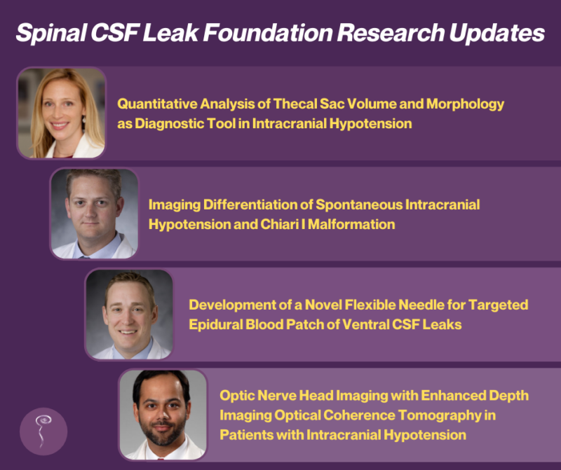
Four grants awarded by the Spinal CSF Leak Foundation are still in progress:
Kerry Knievel, DO, and Lea Alhilali, MD, Barrow Neurological Institute. “Quantitative Analysis of Thecal Sac Volume and Morphology as Diagnostic Tool in Intracranial Hypotension.”
In this study, which is still underway, Dr. Kerry Knievel and Dr. Lea Alhilali of the Barrow Neurological Institute hypothesize that in some patients, spontaneous intracranial hypotension (SIH) “may result more from an increase in the size of the CSF space or increased compliance from connective tissue insufficiency, rather than from significant changes in CSF volume. Detecting these changes in thecal sac volume and compliance in SIH patients could aid in diagnosis in the significant number of patients in which myelography fails to detect a leak, as well as advance our understanding of the underlying pathophysiology.” The study seeks to determine whether or not “differences in thecal sac volume and quantitative shape features (sphericity, convexity, compactness) are sensitive and specific indicators of SIH in patients without detectable leak, improving diagnosis and treatment in this critical SIH population.” Dr. Knievel recently updated the Foundation on the study’s progress so far, noting that while they are still recruiting the remainder of the patients and normal controls to complete the study, on an interim data analysis they were able to find a statistically significant difference in the volume of the thecal sac in patients with SIH compared to normal controls using MRI for volume analysis.
Peter G. Kranz, MD, Duke University Medical Center. “Imaging Differentiation of Spontaneous Intracranial Hypotension and Chiari I Malformation“
This retrospective investigation by Dr. Kranz at Duke University Medical Center aims to determine whether spontaneous intracranial hypotension (SIH) can be distinguished from Chiari 1 malformation (CM1) on brain MRI. This would represent a substantial contribution to care of patients with SIH and CM1 since the ability to distinguish these diagnoses on imaging would be expected to decrease the rate of misdiagnosis and associated inappropriate surgical interventions. Last year, Dr. Kranz was able to update that the study’s data analysis “produced highly statistically significant differences between the groups in a number of different measures, and we anticipate that these differences will be clinically useful in discriminating Chiari 1 patients from SIH patients.” As of now, this study has been completed and is pending review for publication. A poster on this study was recently presented at the 59th Annual Meeting of the American Society of Neuroradiology.
Timothy Amrhein, MD, and Linda Gray Leithe, MD, Duke University Medical Center. “Development of a Novel Flexible Needle for Targeted Epidural Blood Patch of Ventral CSF Leaks.”
Dr. Amrhein initially proposed this study to develop a new flexible needle for epidural blood patching, which could increase the effectiveness and safety of targeting ventral and lateral spinal CSF leaks via epidural blood patch. He recently updated us on his progress:
“Foundation support has allowed for further optimization of this novel patching needle design. New iterations have been tested in cadavers and we are now able to achieve optimal epidurograms (coverage of the ventral epidural space) in 96.8% of needle placements, which is vastly superior to the 47% achievable with standard needles. This work is ongoing and the preliminary data obtained through generous Foundation funding has led to a successful application for a Duke Clinical and Translational Science Institute (CTSI) Translational Accelerator Grant, which will allow for final needle design optimization and initiation of regulatory discussions with FDA in preparation for clinical testing.”
The CTSI grant, awarded in conjunction with the NIH National Center for Advancing Translational Sciences, provides Dr. Amrhein and his team with an additional $123,954 of funding to study “a flexible needle for the delivery of therapeutics in spatially constrained anatomy.”
Fawad A. Khan, MD, and Jonathan D. Nussdorf, MD, Ochsner Clinic Foundation. “Optic Nerve Head Imaging with Enhanced Depth Imaging Optical Coherence Tomography in Patients with Intracranial Hypotension—A Pilot Study.”
As most diagnostic tests for SIH are invasive and often carry significant risks, researchers are interested in establishing more non-invasive methods of identifying SIH. Dr. Khan noted in his comments about this pilot study that since intracranial hypotension can affect the eye, an emerging technique called enhanced depth imaging optical coherence tomography, used in the evaluation of eye diseases, may be one way to test for SIH non-invasively. “This diagnostic test uses ultrasound technology and is not invasive. It allows high‐resolution images of the lamina cribrosa and is superior to magnetic resonance imaging (MRI) and other imaging tests of the eye. Enhanced depth imaging optical coherence tomography is also safer in comparison to the widely used lumbar puncture (spinal tap) in the evaluation of intracranial hypotension. We are committed to investigating the use of enhanced depth imaging optical coherence tomography, a potentially safe and rapid test that can provide data to help in the diagnosis of intracranial hypotension and, possibly, determine the success of therapies.”
Dr. Khan was able to provide the Foundation with the following update: “The COVID-19 restrictions placed on our site during 2020 caused a delay in enrollment for our study. However, we were able to successfully resume enrollment in March 2021, and are slated to complete enrollment by the end of 2021. Our goal is to complete our analysis and present the data at the upcoming neuro-ophthalmology national meeting in early 2022. This will be followed by a formal manuscript detailing the findings.”

