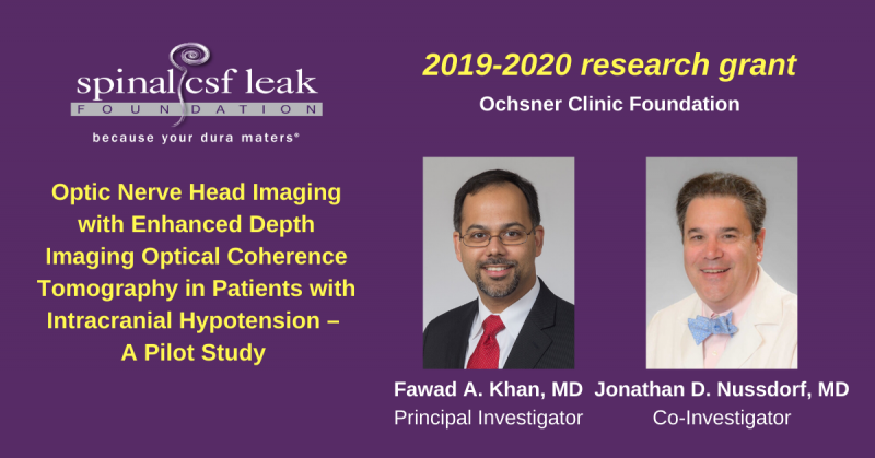
We are pleased to announce this intracranial hypotension research grant to Principal Investigator, Fawad A. Khan, MD and Co-Investigator, Jonathan D. Nussdorf, MD of Ochsner Clinic Foundation in New Orleans, LA. This is one of two research grants awarded during the 2019-2020 cycle, made possible by the generosity of our donors.
Optic Nerve Head Imaging with Enhanced Depth Imaging Optical Coherence Tomography in Patients with Intracranial Hypotension – A Pilot Study
Institution: Ochsner Clinic Foundation, New Orleans, LA
Principal Investigator:
Fawad A. Khan, MD
Director, The McCasland Family Comprehensive Headache Center
Ochsner Neuroscience Institute
Co-Investigator:
Jonathan D. Nussdorf, MD
Chairman, Department of Ophthalmology
Ochsner Clinic Foundation
Intracranial hypotension is characterized by orthostatic headache and a range of other neurological symptoms. Cranial MRI findings are present in 80% of patients, but conversely, cranial MRI imaging may be normal. Cerebrospinal fluid pressure as measured by lumbar puncture may be low but is normal in the majority of patients. Spinal Imaging is normal in about half of patients. Currently, there is no single diagnostic test that either confirms or excludes intracranial hypotension with a high level of accuracy. There is a great need for additional sensitive and specific diagnostic tests in the evaluation of patients presenting with orthostatic headache to help rule in or rule out the diagnosis of intracranial hypotension.
The investigators plan to characterize changes in the optic disc in patients with symptomatic intracranial hypotension, to evaluate the utility of using specific measurements in the diagnosis of intracranial hypotension, as well as the assessment of treatment response.
We asked Dr. Khan to explain the optic nerve head imaging being evaluated in their study in layperson terms.
During intracranial hypotension the brain experiences shifts within the head because of the low-pressure state which can lead to stretching of pain-sensitive structures often leading to headache and neurological symptoms. This shift can extend into other areas directly connected to the brain, for example the eyes. The eyes are connected to the brain via the optic nerve. A structure called lamina cribrosa is situated at the connection point between the optic nerve and the eye. This structure can undergo changes in its shape if the pressure gradient between the pressure in the eye and the pressure in the head changes. If the pressure in the head is elevated the lamina cribrosa can bulge into the eye (for example, a large brain tumor). In contrast, if the pressure in the eye is elevated then the lamina cribrosa can bulge into the optic nerve (for example, glaucoma).
Enhanced depth imaging optical coherence tomography is an emerging technique used in the evaluation of eye diseases by eye specialists. This diagnostic test uses ultrasound technology and is not invasive. It allows high‐resolution images of the lamina cribrosa and is superior to Magnetic resonance imaging (MRI) and other imaging tests of the eye. Enhanced depth imaging optical coherence tomography is also safer in comparison to the widely used lumbar puncture (spinal tap) in the evaluation of intracranial hypotension.
It is not uncommon for patients to go several weeks, and even months, with symptoms related to intracranial hypotension because the standard tests come back negative and the diagnosis remains elusive. Some of the standard tests are invasive and carry significant risks. We are committed to investigating the use of enhanced depth imaging optical coherence tomography, a potentially safe and rapid test that can provide data to help in the diagnosis of intracranial hypotension and, possibly, determine the success of therapies.
We look forward to the study results and will provide an update at the conclusion.
