Many cases of spontaneous intracranial hypotension (SIH) (intracranial hypotension = low pressure inside head) due to spinal CSF (cerebrospinal fluid) leak appear to be related to an underlying weakness of the dura, the connective tissue that holds CSF in around the spinal cord. Heritable (genetic) disorders of connective tissue (HDCT) are associated with spontaneous spinal CSF leaks and are presumed to be a cause of thinner dura. Associated heritable disorders of connective tissue include Marfan syndrome, Ehlers-Danlos syndrome and others.
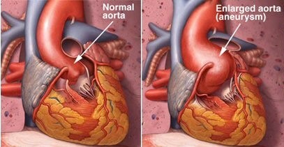
Several HDCT are associated with an increased risk of thoracic aortic aneurysm (enlargement of the aorta in the chest) and other vascular abnormalities. Guidelines for the diagnosis and management of patients with thoracic aortic diseases were published in 2010, including recommendations for screening in patients with HDCT. Depending on the HDCT diagnosis and family history, screening for vascular abnormalities is recommended and may include echocardiography, CT or MR angiography. Thoracic aortic disease is most often clinically silent yet is associated with high risk of death in the case of rupture or dissection. Early diagnosis and management reduces mortality. Patients with Marfan syndrome and some other HDCT are screened with echocardiography which uses ultrasound to image the heart, aorta and other blood vessels. Echocardiography is a non-invasive, low-cost method of screening for dilated aortic root or thoracic aortic aneurysm. Enlargement or dilatation of the aortic root, the first part of the aorta as it exits the heart, occurs early in this disease process.
We have evidence from these associations that connective tissue abnormalities may result in abnormalities of the dura as well as cardiovascular abnormalities. A study was done to look at findings on echocardiography in patients with spontaneous spinal CSF leaks who are often found to have abnormal dura. Patients with spinal CSF leaks from trauma, surgery or other procedures were not studied.
Echocardiographic findings in patients with spontaneous CSF leak.
Pimienta AL, Rimoin DL, Pariani M, Schievink WI, Reinstein E.
J Neurol. 2014 Oct;261(10):1957-60. doi: 10.1007/s00415-014-7438-0. Epub 2014 Jul 25.
Abstract
The presence of cardiovascular abnormalities in patients with spontaneous cerebrospinal fluid (CSF) leaks are not well-documented in the literature, as cardiovascular evaluation is not generally pursued if a patient does not exhibit additional clinical features suggesting an inherited connective tissue disorder. We aimed to assess this association, enrolling a consecutive group of 50 patients referred for spinal CSF leak consultation. Through echocardiographic evaluation and detailed medical history, we estimate that up to 20 % of patients presenting with a spontaneous CSF leak may have some type of cardiovascular abnormality. Further, the increase in prevalence of aortic dilatation in our cohort was statistically significant in comparison to the estimated population prevalence. This supports a clinical basis for echocardiographic screening of these individuals for cardiovascular manifestations that may have otherwise gone unnoticed or evolved into a more severe manifestation.
PMID: 25059392
The authors found that 6 of 50 patients (12%) with spontaneous intracranial hypotension from spinal CSF leak had a dilated aortic root even without a clearly defined heritable disorder of connective tissue. This is higher than the 4.6% reported seen in the Hypertension Genetic Epidemiology study of 2096 hypertensive and 361 normotensive patients in 2001.
Should patients with spontaneous intracranial hypotension have echocardiography? Until we have studied a larger number of patients to better define the prevalence of dilated (enlarged) aortic root, thoracic aortic aneurysm or other abnormalities and whether screening should be done in all SIH patients or just a subset, patients might wish to discuss this study with their physician(s) to decide if they should have an echocardiogram.

