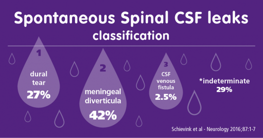
This is a VERY important paper by Schievink and hs colleagues outlining a classification of various types of spontaneous spinal CSF leaks. This does not apply to iatrogenic or traumatic leaks.
The classification was based on a large sample size of well over 500 patients.
Type 1 – Dural tear [27%] – extradural fluid collection in almost all
1a – ventral CSF leaks (95%)
1b – posterolateral CSF leaks (4%)
Type 2 – meningeal diverticula [42%] – extradural CSF collection in 22%
2a – simple diverticula (90.8%)
2b – complex meningeal diverticula/dural ectasia (9.2%)
Type 3 – CSF-venous fistulas [2.5%] – extradural CSF collections absent
Type 4 – Indeterminate [28.7%] – extradural CSF collections were noted in 51.5%
A classification system of spontaneous spinal CSF leaks.
Schievink WI, Maya MM, Jean-Pierre S, Nuño M, Prasad RS, Moser FG.
Neurology. 2016 Jul 20. pii: 10.1212/WNL.0000000000002986. [Epub ahead of print]
Abstract
OBJECTIVE:
Spontaneous spinal CSF leaks cause spontaneous intracranial hypotension but no systematic study of the different types of these CSF leaks has been reported. Based on our experience with spontaneous intracranial hypotension, we propose a classification system of spontaneous spinal CSF leaks.
METHODS:
We reviewed the medical records, radiographic studies, operative notes, and any intraoperative photographs of a group of consecutive patients with spontaneous intracranial hypotension.
RESULTS:
The mean age of the 568 patients (373 [65.7%] women) was 45.7 years. Three types of CSF leak could be identified. Type 1 CSF leaks consisted of a dural tear (151 patients [26.6%]) and these were almost exclusively associated with an extradural CSF collection. Type 1a represented ventral CSF leaks (96%) and type 1b posterolateral CSF leaks (4%). Type 2 CSF leaks consisted of meningeal diverticula (240 patients [42.3%]) and were the source of an extradural CSF collection in 53 of these patients (22.1%). Type 2a represented simple diverticula (90.8%) and type 2b complex meningeal diverticula/dural ectasia (9.2%). Type 3 CSF leaks consisted of direct CSF-venous fistulas (14 patients [2.5%]) and these were not associated with extradural CSF collections. A total of 163 patients (28.7%) had an indeterminate type and extradural CSF collections were noted in 84 (51.5%) of these patients.
CONCLUSIONS:
We identified 3 types of spontaneous spinal CSF leak in this observational study: the dural tear, the meningeal diverticulum, and the CSF-venous fistula. These 3 types and the presence or absence of extradural CSF form the basis of a comprehensive classification system.
PMID: 27440149
DOI: 10.1212/WNL.0000000000002986
