
Sometimes, intracranial hypotension (low intracranial cerebrospinal fluid volume/pressure) due to spinal cerebrospinal fluid (CSF) leak can be mistakenly attributed to Chiari malformation.
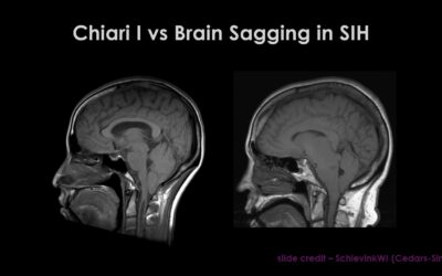
The cerebellum is the part of the brain located behind the top of the brain stem, low at the back of the head. It has two small areas at the bottom of it called the cerebellar tonsils. Normally, the cerebellum and these tonsils sit entirely within the skull. But in Chiari malformation, these cerebellar tonsils—and sometimes the brain stem as well—lie so low that they descend through the large opening on the base of the skull (the foramen magnum) and into the spinal canal. The same imaging findings are also evident in a proportion of patients with intracranial hypotension from a spinal CSF leak, whether that leak is spontaneous, traumatic, or iatrogenic (medically induced).
Chiari malformation is congenital, but descent of the cerebellar tonsils can be secondary to other disorders (also known as “acquired Chiari”), including but not limited to intracranial hypotension from spinal CSF leak. There are cases of acquired Chiari that are the result of other conditions, such as a brain tumor or intracranial hypertension.
Often the symptoms people experience with Chiari malformation are similar to those experienced by people with spinal CSF leak—such as pain in the occipital (back) part of head or suboccipital (lower back part of head), or headache that worsens with cough. Other similar symptoms include neck pain, imbalance, dizziness, and limb numbness.
Despite the similarity of symptoms, there is a big difference in treatment: in cases of true Chiari I, surgical decompression is sometimes recommended. But if the brain descent is actually secondary to loss of CSF volume, then correction of the underlying cause (treating the spinal CSF leak) will usually result in return of the brain to its normal position.
If Chiari I is seen on imaging, usually MRI, then reversible causes such as intracranial hypotension from spinal CSF leaking or intracranial hypertension must be ruled out. In cases of intracranial hypotension, additional imaging findings will usually be present, which will help with correct diagnosis. Additional imaging findings on cranial MRI in cases of intracranial hypotension include:
S = subdural fluid collections
E = enhancement of pachymeninges
E = engorgement of venous structures
P = pituitary hyperemia
S = sagging of brain (there is measurable mid-brain descent in addition to low-lying cerebellar tonsils in Intracranial hypotension)
Remember, low-lying cerebellar tonsils can be congenital or secondary to another, often correctable, cause. The underlying cause of intracranial hypotension due to spinal CSF leak should be considered so that the most appropriate treatment can be planned.