
Chiari 1 Malformation and Spinal CSF Leak
Sometimes, intracranial hypotension (low volume of cerebrospinal fluid within the skull) due to spinal cerebrospinal fluid (CSF) leak can be mistakenly attributed to Chiari 1 malformation.
What is Chiari 1 malformation?
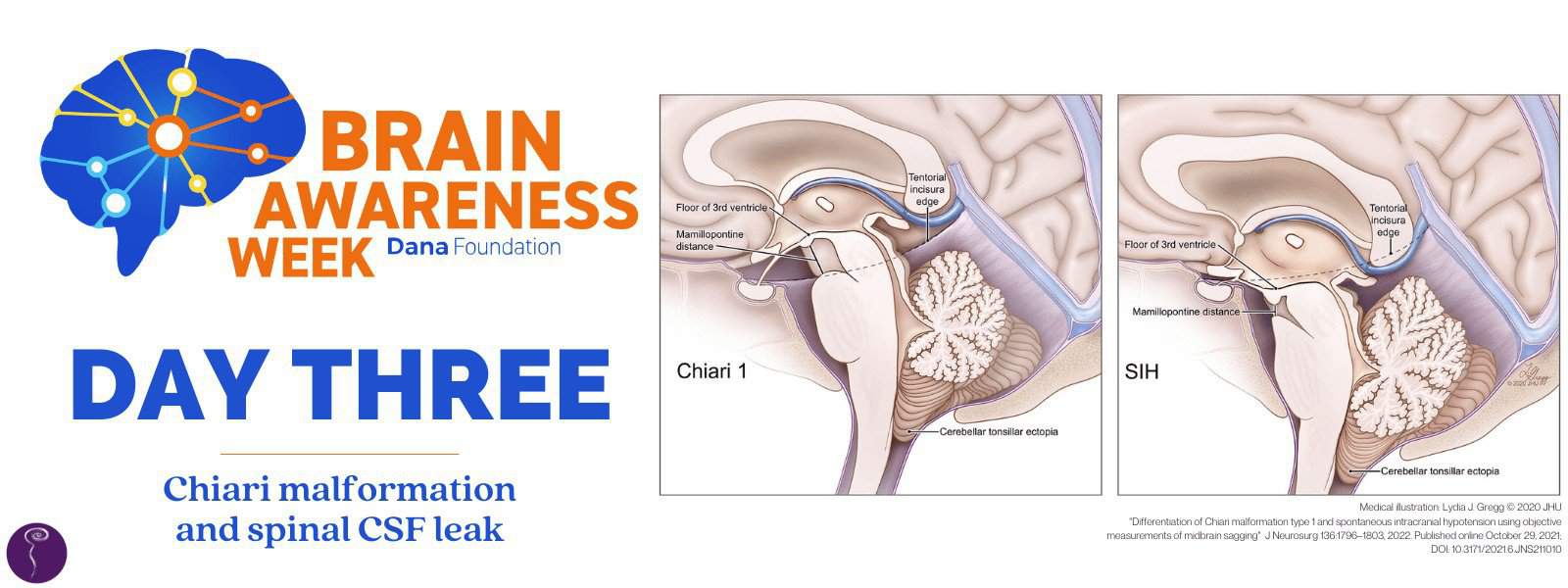
Chiari 1 malformation is a congenital structural defect in the base of the skull and cerebellum. The cerebellum is the part of the brain located behind the top of the brain stem, low at the back of the head. It has two small areas at the bottom of it called the cerebellar tonsils. Normally, the cerebellum and these tonsils sit entirely within the skull. But in Chiari 1 malformation, these cerebellar tonsils—and sometimes the brain stem as well—lie so low that they descend through the large opening on the base of the skull (the foramen magnum) and into the spinal canal. This is called cerebellar tonsillar ectopia.
How does Chiari 1 differ from spinal CSF leak?
The same imaging findings of “brain sag” and cerebellar tonsillar ectopia are also evident in a proportion of patients with intracranial hypotension from a spinal CSF leak, whether that leak is spontaneous, traumatic, or iatrogenic (medically induced).
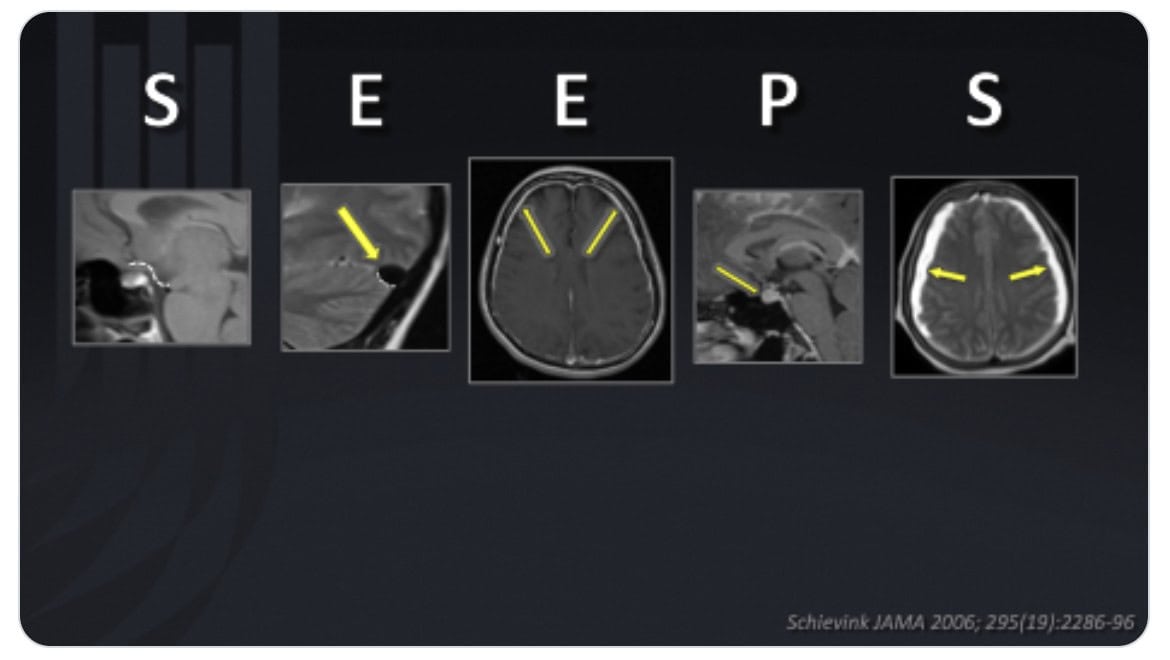
However, in cases of intracranial hypotension, additional imaging findings on cranial MRI will usually be present. These additional imaging findings, outlined here using a mnemonic device coined by Dr. Wouter Schievink, include:
S = subdural fluid collections
E = enhancement of pachymeninges
E = engorgement of venous structures
P = pituitary hyperemia
S = sagging of brain (there is measurable mid-brain descent in addition to low-lying cerebellar tonsils in Intracranial hypotension)
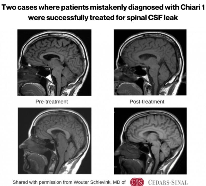
This is important to note, because there is a big difference in treatment between Chiari 1 and spinal CSF leak. In cases of true Chiari 1, surgical decompression is sometimes recommended. This surgical intervention may improve Chiari 1, but will do nothing to resolve a spinal CSF leak (and may even make it worse). However, if the brain descent is identified as being secondary to loss of CSF volume due to a spinal CSF leak, then treating the spinal CSF leak with less invasive procedures will usually result in return of the brain to its normal position.
A study on Chiari 1 vs SIH
A study by published in the Journal of Neurosurgery Dr. Peter Kranz at Duke University Medical Center titled “Differentiation of Chiari malformation type 1 and spontaneous intracranial hypotension using objective measurements of midbrain sagging” demonstrated that there are important differences between Chiari 1 and spinal CSF leak that can be seen on brain MRI.
This retrospective study, funded by the Spinal CSF Leak Foundation, compared neuroimaging in adults with Chiari 1 and those with intracranial hypotension due to spinal CSF leak. The results showed highly significant differences between the groups. Importantly, it also showed that none of the patients with spinal CSF leak had cerebellar tonsillar ectopia without also having other findings of midbrain sagging. Ultimately, the study demonstrated the importance of looking at objective measures of midbrain sagging to help prevent misdiagnosis and unnecessary surgery.
Conclusion
Though there are similarities in some of the symptoms and imaging findings between Chiari 1 and spinal CSF leak, there are many important differences, and misdiagnosis can lead to unnecessary surgeries and prolonged suffering for spinal CSF leak patients. An underlying cause of intracranial hypotension due to spinal CSF leak should always be considered so that the most appropriate treatment can be planned.
Further reading:
For more information on Chiari Malformation, visit Bobby Jones Chiari & Syringomyelia Foundation
Chiari Malformation Awareness Month