
Slides
Transcript
Yeah, good morning. Thank you. This is a wonderful work you’ve presented, Tim, and so much work to analyze all the data from the literature. Congratulations.
So what I want to bring up to the stage is what shall we use — surgical ligation, endovascular embolization, or percutaneous procedures? First of all, of course, once again, the thing is not only which method we should choose, but also that we really treat these patients. And again, quality of life — you mentioned it as severe as end-stage cancer patients, HIV patients — so really do not forget this.
So still, I think one of the biggest game changers could be that we raise awareness with the general practitioners and neurologists to treat these patients. What to treat and how to treat — I cannot add anything more to this discussion. I don’t find another RCT that hasn’t been described yet. It’s really so subjective how to analyze the literature, and Levi and Tim have really shown that the outcome parameter is getting better is so subjective. It’s done the next day after blood patching, it’s done a few months after blood patching, five years. Nobody really knows what happened to the SLEC.
So I cannot really add anything significant to this discussion. I show you the videos in the end only to present our work, our results. For instance, these are the results for surgical intervention, the nerve root clipping discussion for fistulas. One parameter that might be a little bit more objective is return to work. In this series, I think there are 60 patients or so. None of the patients — all 30 to 60 years, in the middle of their life — were able to work. That’s reached our selection bias and so on service. After surgery, after nerve root clipping, 80% were fully employed. Only 11% were left not able to work 100%. Most of them returned to work, but not 100%. But this is, I think, an extremely good result based on an objective outcome parameter — return to work.
This is a larger series. Florian Volz is currently working up and trying to publish over 200 patients including all etiologies — CSF-venous fistulas, ventral leaks, lateral leaks. Again, we can see how severe the disease is affecting our patients. Before SIH, these over 200 patients all were in the middle of their life working. After the diagnosis, two-thirds were not able to work, and the ones that were able to work, only 5% or so were able to work full-time.
Three months after surgery, dramatic improvement, but still a third were not able to work. And despite we analyze our patients currently only tried on objective criteria — meaning is SLEC gone, can we find, do you repeat imaging if you have a fistula — so really objective criteria. And even two years after surgery it continues to get better, but I think 15% or 20% or so are not able to work.
So this is, on the one side, pinpointing to how we should assess treatment access. I think imaging-based is fistula gone, and still we’ve discussed this extensively yesterday. If you treat late, if you have a patient with years of suffering and symptoms, despite closing — and this is so hard to get for us, closing something, a hole, you can suture, you can close — still the patient does not get better. And this is kind of long SIH. So we need probably to find a good term for that. It has probably nothing to do with the effectiveness of our procedure, nothing to do with the effectiveness of blood patching, nothing to do with the effectiveness of surgery, but with the cause of the disease and with the changes the longstanding spinal leak has induced in these patients. We need to add different treatments plus patching or specific treatment to really heal our patients.
Again the effect of surgery is dramatic. A delta of 15 — five times the delta that is required for all this expensive migraine medication. To add a little bit to the confusion, this is just, I think, one of the recent papers on how good endovascular procedures are as compared to surgery. Here now for fistulas, complete improvement in the Brinjikji Mayo series, 58%. Time again was the only modifiable treatment risk factor. And the excellent series from Andy and Lalani also complete improvement, 60%.
This is not comparable now, but again another measure is this is our series for the surgical intervention for fistulas with nerve root clipping, and this is done by the patient global impression of change outcomes. So PROMs — patient-related outcome measures — that the patients are asked for via email, standardized in Freiburg, 3 months, 6 months, and one year later. There were 98% improvement, 85% very much or much improved. So this is not comparable, but I think when we rely more on objective parameters, targeted surgical treatment is clearly the best way to proceed and clearly superior to endovascular, to patching, to all the non-surgical treatments.
And the best evidence for it, if you really look very, very, very closely to patients that really do suffer, as Wouter did in his superficial siderosis series. This is a meticulously controlled study now because we do have this horrible sequelae of superficial siderosis we discussed yesterday. All these patients — I have forgotten, I think 50 or so — have been followed up with imaging — what happens to the siderosis, what happens to the SLEC? And all of them, all of them — what better study can you have? All of them have been patched, and no single patch closed the leak. But all of the surgeries closed the leak, at least the ones where we found the leak, where the site was known, and surgical repair was associated with a very low risk. So what do you want more? This is the proof that surgery works and blood patching doesn’t work. Period.
Why? You know that it cannot work even in the best hands. I like the study when Andy and Lalani put needles in CT-super-controlled fashion into the membrane — still only, I think, 20% to 25% sealing of the leak. So again, there’s the proof. We have the best studies so far. Now you know why — because there are membranes. This was also discussed yesterday.
Just to give five minutes very provocative input: blood patching does not work at all. Go for surgery. Thanks.
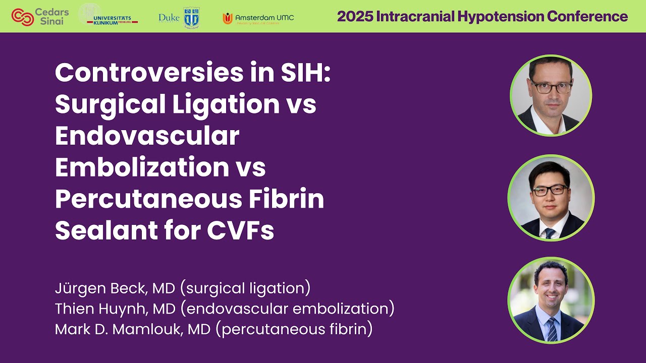
Controversies in SIH: Dr. Thien Huynh on Endovascular Embolization
Thank you very much. It’s quite an honor to be here in front of all the Michael Jordans of the field, who I basically read the papers like the gospel for the past several years. So it’s just quite an honor to be here.
I get to talk about something very near and dear to my heart that I’ve been doing for the past 5 years here, which is transvenous embolization of CSF-venous fistulas. I’m going to be focusing on the fistulas. I don’t think anybody’s onyxing type 1 leaks hopefully.
But basically these are the kind of success stories that we see now in my practice. As an interventionalist who treats stroke, I mean the work we’re doing here is equal or more impactful. So it was quite surprising to see patients presenting with the frontotemporal dementia syndrome, but now looking at this MRI pretty much everybody in this room knows what to do.
In my shop parts, I personally I do enjoy CT myelograms. As Lal talked about, I have a certain bolus tracking technique where I can monitor the bolus and make sure it’s right there and take a snapshot, and we can see beautifully the enhancing paraspinal veins. This is what an embolization cast looks like. It’s very bizarre looking to begin with, but once you get the hang of it and you start to see good outcomes, you can really appreciate that that’s what they’re supposed to look like.
This is what the patient looked like before and shortly after, within the month or two months, they had complete resolution of their dementia and headache within the month, and their MRI brain looks fantastic. So I think we’re starting to reach the golden age, to be honest, in this SIH leak work, and especially as neurointerventionalists get involved, it’s quite a privilege and honor to be involved.
So, my point here today is why do we treat these endovascularly versus fibrin or glue or surgery? I used to be a very pro-fibrin guy. That’s basically all we had as neurointerventionalists before, and unless you knew a Michael Jordan, Dr. Schievink, Dr. Beck on board who you could call in, then you were kind of SOL.
This was when my first patient we struggled with, and they had the CSF-venous fistula at C7. We tried patching it quite extensively, and every time one to two months it would come back. I’ll tell you, these are hard work to get it right there perfectly at C7. This was when I first started doing fibrin patches, I would not recommend doing that interlaminar thing I did on the second slide there. But and then it would work, but then in one to two months it would come back. It was like as if you never did it.
Then in particular, this was a C7 case. I’m not sure exactly how hard it is to preserve that root, but I can’t imagine that would be a slam dunk thing as well. Luckily, Waleed sent an email right exactly at that time in November 2020 — very appropriate title, “The Next Frontier: CSF Leak Treatment.” In that, he really outlined that if you could get there with a vein as he was seeing them light up, if we can navigate there, wouldn’t it make sense to embolize them?
Because one, instead of the fibrin dissolving, the embolic agent is confined to the vein and it permanently occludes it, so you can guarantee that it’s going to stay. Number two, it’s minimally invasive. It avoids an open surgery, and it potentially could preserve nerve roots at eloquent levels. And it’s the safety of a transvenous procedure. Nothing is more delightful than a transvenous procedure for a neurointerventionalist.
So that’s what his first cast looked like, and it was very quite elegant. To my amazement, all his patients — his first five patients — reported basically 100% partial or complete improvement, and all had significant improvement in their brain MRI findings. So definitely we’re on to something.
This is where the first case that I had was, and it was at C7. It’s quite daunting. This wasn’t his standard case, and this is what I had. He basically called me and said, “This is what you’re supposed to do,” but I kind of did the whole thing on my own. Once you start to see veins like that, you kind of have to take a pause and think, “What am I doing here?” But what the point is that we’re trying to achieve the same thing that surgeons can do, or fibrin patching. We’re trying to occlude all the veins in the tributaries around the CSF-venous fistula. So if we can get right there and prove it because we have our anatomical imaging, then we can just glue them up permanently and we should be done.
That’s what I did for that patient. To my surprise, unlike the fibrin patches which dissolved, this one stayed for six months. This patient I first did in 2020, and they’re still solid now for about four or five years now. So this has really changed my thinking about the fibrin glue.
This is another case that was done before my time. I started in 2020. You can see my colleagues before were again trying to do everything they could. They had a leak at T4. I’m sorry to say that wasn’t lateral decubitis myelogram, but they still suspected something real there. They patched it twice. Patient got an improvement maybe for a month or two, came back. You can see their first leak sign was in 2016. Now this is ongoing till 2020. And then finally we did a DSM, proved the site. This was actually on the left, the T4, and you can actually see the superior supreme intercostal vein on that left side, the aortic nipple coming across the aorta. If you can just get into that vein and do a microcath injection, prove you’re right in the neuroforamen, then you’re golden. And so that’s what we did. We embolized this quite thoroughly, trying to do again what the surgeons are trying to do, and then we could see within 3 months the whole brain start to lift up, again achieving something that we could only dream of with the fibrin glue.
So CSF-venous fistula embolization was very impactful and very rewarding if done correctly. And I should say we’re starting to see some issues, which I’ll get into. At the time, for the next few patients we had a real discussion with them, a frank discussion. We have a new treatment, embolization. It seems very good. It seems to be a little bit better than our previous experience with fibrin patching. We know fibrin patching may be very variable. This was kind of before the Mark Mamlouk days of 100% — things like that. And it was very variable. Maybe if you live close and you don’t mind getting a couple patches it’s not a big deal, but patients don’t necessarily want to hear that.
The only nice thing about fibrin patching is it doesn’t leave any residual dense artifact, which is very nice. So now compared to surgical ligation, that’s more of a slam dunk thing that may work, but it’s a little bit more invasive. You might require a laminotomy. You may or may not require the nerve root to be clipped or sacrificed. The Adamkiewicz, you know, is probably not a big deal, but we don’t really know. And then, locally, if you’ve got a Michael Jordan ready to go, then by all means, but many of us struggle for that.
So ultimately the conversation led to that really most patients want the high efficacy treatment with the least morbidity with the single treatment. So that really kind of allowed CSF-venous fistula, at my institution at least, to take off and across Mayo really. So now the procedure is performed internationally. Mayo Rochester has done over 400, we’re about 100, and it’s validated in multiple international cohorts. Most recently I got to thank Frederico for his nice papers in INR, which the French group is catching up. So please, please, everybody get your publications out there because I know a lot more are being done.
Overall the summation is that there’s about 90–94% clinical improvement, at least partial or complete, and 80–90% imaging improvement with the mean follow-up of 15 months.
This is just an example of what I saw in my experience in our 18 patients. You can see many of those blind [inaudible] down to no symptoms at all and significant improvements in the Bern score, which was terribly exciting.
Another interesting thing is that when we start to image these patients soon after treatment, we could actually see that the brain was starting to lift up in some patients pretty quick, within 24–48 hours. We published this in Neurology. You can see the suprasellar distance improved, the mammillopontine distance improved very quickly. The venous distension improved as well as a little arachnoid granulation even showing up.
This was replicated in Dr. Cagnazzo’s reports in about 40 patients. And here you can see the C image is the 24 hours and the D image is the 3-month scan. You can see the pituitary again gradually improving. He actually found that pachymeningeal enhancement goes away pretty quick. So that again confirms the efficacy both short-term and long-term of transvenous embolization in a larger cohort.
Recently Dr. Kallmes et al. sent a nice paper to AJNR comparing a systematic review between embolization and surgical treatment for CSF-venous fistulas. Their ultimate conclusions were similar — partial or complete response rate over 90% for both. Symptom resolution was also very similar. They found it slightly lower, 60% versus 59%, 16% for embo versus 70%, which is not significant, and similar retreatment rate. So I think we’re getting very good results compared to the surgeons. Here’s just an example of their chart showing the partial or complete response rates well in that 80–90% for both.
Another thing is it’s very safe. In our series at Mayo, we had 100% technical success rate, no neurological complications to the cord or regarding cord compression or intrathecal extension. I did have one case of radiculopathy that persisted, but other than that, at the T1 level, we had a few cases of pulmonary emboli which were asymptomatic, and we did perforate some epidural veins, but again that didn’t have any clinical sequelae. You can even consider that an additional blood patch there actually.
But there are issues though. The main thing that we all need to think about is these treatment failures that seem to pop up. The real tricky thing here is that essentially what we’re trying to treat is almost like a dural fistula that we can’t even confirm where it is in the procedure or whether we closed it during the procedure.
So it’s like as if you’re treating a spinal dural fistula, cranial dural fistula, with just Onyx without being able to confirm. And so the only way you can know if you got it is by spatially locating where you think the leak is and nailing all the tributaries in case you don’t hit the spot. There are some nuances to it. It’s not simply like an MMA embo where you can just throw something in there and call it a day. This is something where you really have to put your neurointerventional hat on and be an artist.
Having said that, to be an artist in this, you do really need great pictures. Again, these are the pictures that we’re getting with CT myelograms, where we can actually see all those tributaries, and all the nice review papers that have been published. Here you can see that from that one level it’s going into the paraspinal vein, but wait — it’s going into the dorsal muscular branch, intervertebral venous plexus, basivertebral vein. And if you don’t nail each one of those, then obviously the CSF can leak around your Onyx. I mean, if you ever held Onyx in your hand, it’s like the water just flows right by it. So it’s not something that will be inflammatory and occlude everything on its own. Again, this anatomy that we’re seeing pretty much matches what the Duke team published here. It looks almost identical. Again, those are all the little tributaries that you have to know the names of and occlude.
This is one of the cases that taught me the importance of looking for all these little branches. So the one on the left case, to all eyes looks pretty good on the AP, but on the lateral you’re actually missing maybe something in the dorsal muscular branch area and around the posterior aspect of the neuroforamen. This patient actually didn’t improve despite a decent looking cast. What I found is you really, really have to be very thorough when you embolize these, including getting those dorsal muscular branches as radiologists have always seen.
These are some things that we’re learning, and I’m having very, very nice results thinking about these concepts. Being sure to thoroughly embolize everything, we came up with this dual microcatheter and balloon pressure cooker technique. The concept here is you have your leak at the foraminal venous plexus and then the CSF is basically flowing out from there, and it can go in any which direction. Not only that, it’s going downstream, where your Onyx has to go upstream.
So basically you have to put little coils to plug where the Onyx shouldn’t go and put balloons there, using the pedicle as a marker. You get one microcatheter kind of under the pedicle and the epidural venous plexus and the balloon where you like, and then basically just embolize everything and be very thorough to build Onyx plugs as you would for a dural fistula. Again, you’re fighting against the flow of the CSF, so it’s not going to naturally go there, and plus the Onyx will go to the big things first. This is just an example of a cast. I wish I coiled up that little coil to make it bunch up a little better, but it is what it is.
This is also effective not for the simple fistulas but for complex fistulas. So this is a very interesting case of a 50-year-old woman. Her main issue was orthostatic headaches, neck pain, as well as progressive upper extremity and lower extremity weakness, myelopathy, and numbness. This was progressive — she was going to the ED. You can see there’s a massive, massive syrinx here associated with the altered cranial flow at the craniocervical junction due to CSF leak, described by my colleague Eric Middlebrooks.
And so this was her myelogram. I use this as a great example to see everything you need to get. Again, you want to nail this on the first time. You don’t want to have to come back and worry about repeating myelograms. Plus, she’s getting worsening myelopathy, so the stakes are high. This is what her embolization looks like. We were very, very thorough, as we hope. And then, 3-month follow-up, her syrinx was totally gone and her myelopathy halted. You can see the nice raise in her brain. And that’s been good now for several years.
Regarding incomplete embolization, I was very impressed with Dr. Schievink’s paper, as always, about surgery being a viable option after embolization. I think they had six cases that failed. I was initially shocked, because although the surgical technique was beautiful, I didn’t have six failures per se due to that. So I think the technique does make a difference, but good to know that if you’ve got a Michael Jordan around, they can save the day.
Another interesting phenomenon we found with these leaks is that some cases that don’t seem to work, it could be that, as Lal pointed out before, you’ve popped out another level. Again, I’d love to know the pathophysiology behind this — what is happening here. But clearly, we can see that you can pop a new level that was not there before the first time. This happens with fibrin glue. This happens with surgery. This happens with embolization.
I know I’ve taken up a lot of time here, but I do want to share this beautiful case I had to this great audience. So this is the pièce de résistance for me as far as it goes for transvenous embolization CSF leak. We have a 30-year-old lady with classic intracranial hypotension. You can see their brain sag from a mile away. Just like many patients — they’ve got their lateral decubitis DSMs. They even had a positive pressure MR myelo because we were desperate. We always saw renal contrast excretion, but we never actually saw the leak site. She’s had multiple epidural blood patches, as we talked about, fibrin patches without improvement, persistent proven clinical imaging findings. This is just a finding of what we had.
We repeated it at our center just to make sure, because sometimes these things want to show up one day or the other. They always had renal contrast excretion. The left side was negative, the right side showed this extensive contrast leak prior to the right side injection. Hard to know what that was from.
We repeated the left lateral injection just to see what was going on because the contrast injection in the kidney was so remarkable. Just as I was about to sign off the study saying it was negative, I realized I scanned quite high on one of them. I just looked there, and lo and behold, I just looked for any contrast density outside the spine and I saw something in the paraspinal veins right at the C1–C2 level, which we really don’t ever look very high that often. But I’m glad we did.
It looked like a fistula at this level. We brought them back, did a dedicated study looking at that right area, and it actually proved to be a hypoglossal canal CSF-venous fistula, right at the anterior condylar confluence, which I don’t think has been reported yet. So we show nicely in this coronal MIP image here where there’s a diverticulum and the CSF-venous fistula coming out. This was a very special case because it’s cool enough to show the fistula, but now we actually have to fix the patient. Fibrin glue — I don’t know if the Mamlouk’s of the world can cure this. I’d love to see them try, but surgery I think would be very difficult here. So, transvenous embolization is very natural.
We mapped it out all in the T2-space MRV to see exactly what the tributary veins were. Here’s the diverticulum. Here’s the CSF flow coming out. We can overlay the images to see exactly what we need to occlude, and again this brings it back to beautiful anatomy that as neurointerventionists we love. So we basically have to glue up all the tributaries of the anterior condylar confluence and the hypoglossal canal. We navigate there with a catheter, do a run, and now we can see this beautiful anatomy exactly around the leak site. I won’t belabor it, but basically we use the balloon catheter technique where we can occlude the Onyx from going into our jugular and make sure it’s confined right to the hypoglossal canal using the pressure cooker type of technique. This is the microcatheter injection right before the Onyx, and then this is our balloon is inflated — this is the final Onyx cast going into the hypoglossal canal. Here’s the Onyx cast. You can see mainly the coils and Onyx in the suboccipital venous plexus, then coming up to the anterior condylar confluence and then going into the hypoglossal canal exactly like we like — just to cup that diverticulum basically.
The patient initially developed rebound headache. They did have mild tongue deviation for a little bit, but that resolved, and then basically by two to five months their imaging markedly improved and clinically they’ve gone back to their work-life baseline, and they’re very much improved on the PGIC scale. Just wonderful to see, and then here’s an interesting distension of their optic nerve sheath again. Interestingly, did have some venous sinus stenosis, which we did treat with Diamox, and she’s controlled with that.
With that, I’ll conclude. Basically, as the Michael Jordans say, “Patients and their healthcare providers are fortunate that the armamentarium for treating spontaneous CSF leaks — CSF-venous fistulas — is expanding.” It’s very highly efficacious and safe. I think we can consider it durable first-line treatment for CSF-venous fistulas. I think comparable to surgical ligation in the right hands and probably maybe a little bit more reliable to fibrin glue in the right hands. Hard to say, but I think these things are all highly operator dependent. There’s a lot of technical nuances, which I went through. You do have to be an artist about it, and there are a lot of opportunities for further research in this new frontier to improve patient care. Thank you very much.
Controversies in SIH: Dr. Mark Mamlouk on Percutaneous Fibrin Sealant
Thank you for that kind introduction. I’d like to thank Dr. Schievink for the invitation to speak and our hosts here in Amsterdam for their wonderful hospitality.
So in this talk, I wanted to share our experience performing fibrin glue patching in our first 100 patients. Before I get into that, I want to share an index case of a recent patient with headaches who presented to the ICU for subdural hematomas and brain sag.
This is day one of the admission. The very next day of the admission, we did a myelogram. We identified a right T6 fistula. In the same session as the myelogram, we performed a fibrin glue patch. This is day two. Day three, the patient felt better and was discharged. At a later outpatient date, we did a follow-up MRI showing resolution. This case illustrates the ease and efficiency of fibrin glue patching. What would have previously taken maybe weeks to months to years, now we can make a diagnosis and treatment in just a few days. I think that is really one of the advantages of fibrin glue.
So in this talk, we’re going to go on a spinal leak journey, and we’re going to have three stops along the way. We’re going to talk about how to perform a fibrin glue patch, some of the tips we’ve observed that can lead to a successful patch, and lastly close out with a patient story about additional fibrin glue patching opportunities.
There are five steps to perform a fibrin glue patch. The first step is to scan prone on CT at the fistula level. Now this can be performed in the same session as the myelogram or a different session, although we prefer to do it in the same session because having that meningeal diverticulum opacified can really help with needle placement. Speaking of needle placement, we use one or two 20-gauge needles and we place it along the fistula course, and I’ll talk about where to place that.
We do a generous test dose with air or iodine. We inject contrast into both hubs of the glue, and then lastly we inject 1 to 2 cc’s. We do a quick scan check and 1 or 2 cc’s more. All patients receive conscious sedation. If patients have received fibrin glue previously, we administer Benadryl.
Here are just a few videos of the fibrin glue prep and the injection. The fibrin glue comes frozen, so we place it in a warm bath to thaw it. The second image, the second video, shows our tray, and then you’ll see the fibrin glue. When we’re ready to inject, we pull back on the plunger, inject a little bit of contrast into both hubs of the glue, give it a little swirl to kind of mix up the contrast and the glue. And then lastly, we’re ready to inject. So we take out the needle stylet, connect it, and then inject 1 to 2 cc’s, put the stylet back in, do a quick scan check, and repeat as necessary.
So what are the tips of success? We observed three tips that have led to success. First tip: location, location, location. There are unique places and specific places you want to inject, and other places that may not be as helpful. The second point is glue spread matters. You want to inject the glue where the fistula drainage pattern occurs. I can’t harp on these two points enough. The last point is extrinsic compression is probably better than intravascular coating of the glue. When we first started doing this, we thought that the trick or a bonus was intravascular glue. And while that may be helpful in certain scenarios, now we like to think that the mass effect are the best ways to keep that vein nonfunctional.
So where exactly do you inject? Here is a decubitus myelogram. You can observe the meningeal diverticulum and there’s a vein. The first area that we like to inject is at the cyst-vein junction. One of the goals is to sever the connection between the cyst and the vein. The second area is this paravertebral wall. We like using this because injecting in this location and using the vertebral body as an anchor, glue in this location can then compress the vein against the bone and make it nonfunctional.
Lastly, if there’s no ways to access the veins, then we can do a direct cyst puncture and there are a few indications for this. Now, note the fistula in this case. If you did an epidural interlaminar patch, the epidural space here would be wasted glue. So, it’s actually a misnomer — epidural patching for CSF-venous fistulas — because if there is no epidural involvement of the fistula, then glue or blood in this location will not have any benefit. So you really want to target your glue with where the vein is.
So let’s go over a few examples. The first is targeting the cyst-vein junction. This is a patient with SIH, had a decubitus myelogram, and had a couple of veins connecting to that cyst and then a basivertebral vein. We had two needles in this slow case. We’re advancing and advancing and advancing. The first needle is in proximity to one of the veins and then the second needle is in proximity to the second vein. We do a generous test dose and then inject glue. Inject a little bit more glue. Here’s a cine clip that also demonstrates that. Notice that the glue is coating the cyst-vein junction. We did a follow-up myelogram. We don’t typically always do this anymore, but this is one of our first cases, and we can see resolution of the fistula. Follow-up MRI — resolved. Patient’s symptoms clinically resolved.
I really like this next case. It’s an unusual fistula starting at left T9 and extending cranially in the internal epidural plexus, exiting at the left T8 level. You’ll see this huge segmental vein going into the hemiazygos.
In this particular example, we did two needles, but at two different levels. One at the origin at this left T9 level and then at a level higher at the left T8 level. Here’s the initial MRI showing brain sag and subdurals. At one month, the brain sag resolved, the right subdural resolved, and the left subdural markedly decreased.
Here is an example of the paravertebral wall technique. I really like this technique. Here’s a patient with SIH and a left T8 fistula. We also used two needles in this particular example. We’re advancing to the paravertebral space. There’s one needle, there’s the second needle. We do an air test. We inject — inject a little bit more, and you can observe a nice wall coating of that glue and then also inject it in the second needle. Here at one month showing complete resolution, patient resolved.
Now one thing about fibrin glue patching — one of the things to consider is that it may take more than one treatment to be successful. What we’ve observed is that real-time adjustment of the needle can decrease the number of attempts.
Here was a right T6 fistula, and we placed our needle at the cyst-vein junction. I was very happy with the coating here, but I wanted to be extra confident. So we then advanced our needle a little bit more eventually, and you can observe glue within this paravertebral space. So we have glue in this paravertebral wall as well as at the foramen, and this patient went on to have a cure.
So using this real-time needle adjustment can help you decrease the number of attempts. Last location that I’ll share is this direct cyst puncture. Here was an unusual fistula. You observe a large cyst here on the left T10 level and then there’s an internal epidural plexus vein, and then you’ll see drainage on the contralateral side. In this particular example, on the first attempt we just targeted the right side, although that wasn’t the origin, and it wasn’t successful. So we brought the patient back and then did a direct cyst puncture. Here is the air test and then here is glue, and then also did a second needle on the right side. Here is the pre-treatment MRI showing diffuse dural enhancement, and at the follow-up MRI there was near complete resolution of the dural enhancement. So the direct cyst puncture may be used in certain scenarios.
How good are the data for fibrin glue patching? I think the largest study to date is this multi-institutional and multi-country study. 60% of patients had complete improvement, 35% partial, and 6% had no improvement. As mentioned before, glue spread matching the fistula drainage pattern was a statistically significant variable. This corresponds to a recent study from Dr. Cagnazzo’s group on embolization where they looked at several metrics, but one of the metrics they looked at was headache response, and 64% had complete headache response. So very similar data from embolization and fibrin glue.
Lastly, I just want to share one final case that really illustrates one advantage of fibrin glue patching. It was a recent case that I encountered. I’m a volunteer professor at University of California, San Francisco, and it was a case I was just reading with the residents. It was a 23-year-old woman who presented with headaches from neurosurgery for Chiari evaluation. You can observe there is cerebellar tonsillar descent, there is a syrinx, however there is brain sag as well, and as many of you know that’s not really consistent with Chiari. I wondered if there was SIH here, but 23 years old is actually very young for most SIH patients.
I looked a little bit more in the chart and saw this outside spine MRI, and there was this very bizarre dural ectasia. I said that’s really interesting and looked a little bit more, and then on the sagittal MRI there was this paraspinal lesion that I don’t think was even mentioned on the outside report. I told myself that’s really interesting. It got more interesting. The patient, when she was one year old, she had this MRI and there was this enhancing lesion at the T12 level, same exact location. At the time it was read out as epidural hematoma or hemangioma.
So putting it all together, I wondered if this represented SIH from a venous malformation fistula and suggested she come back and do a right decubitus myelogram. Sure enough, we did just that. And here’s the early myelographic phase. You can again observe this very bizarre dural ectasia, but we didn’t observe any fistula. We did see a phlebolith, so we knew we were in the ballpark of a venous malformation. So we simply then just did a delayed scan. And now you can observe abnormal connection between the subarachnoid space and the lesion, consistent with a CSF-venous malformation fistula.
At this point I was actually really happy and ready to call it a day. But my colleague Bill Dillon asked me if I wanted to treat it, and frankly I told him I’ve never treated a CSF-venous malformation fistula. But sure enough we did, and we injected glue exactly at the location of the fistula’s connection. At a later date, one of my colleagues did a sclerotherapy of the venous malformation, which we subsequently published.
So hopefully in this talk I’ve shared with you some of our experiences of how to perform a fibrin glue patch, some of the tips for success, and lastly some additional image indications. And with that I’d like to thank you for your attention.