


This is Chiari Awareness Month.
What is Chiari malformation?
In Chiari malformation, the cerebellar tonsils (part of brain) are displaced downwards below the opening at the base of the skull. Up to 3 mm of descent is considered within normal limits; 3-6 mm of descent may or may not be normal; 6 or more mm of descent is considered abnormal. In some cases, this will not cause symptoms, but in many, symptoms can be quite significant. Surgical decompression is sometimes recommended.
Chiari is often, but not always, congenital. There are cases of ACQUIRED Chiari that are the result of other conditions, such as a brain tumor. The same imaging findings are ALSO evident in a proportion of patients with intracranial hypotension (low cerebrospinal fluid or CSF volume) from a spinal CSF leak. This can be seen in patients with spontaneous, traumatic or iatrogenic (medically induced) spinal CSF leaks. Learn more about causes of spinal CSF leaks here: spinalcsfleak.org/?p=34
The most common symptom in patients with Chiari is headache with the location in the occipital (back of head) region. The headache from low CSF volume (intracranial hypotension) is most often in the same location. Other symptoms in patients with Chiari or patients with spinal CSF leaks are also similar such as neck pain, imbalance, dizziness, limb numbness.
Why is this important?
If the brain descent is secondary to loss of CSF volume, then correction of the underlying cause (treating the CSF leak) will usually result in return of the brain to its normal position. Patients with spinal CSF leaks and acquired Chiari on imaging RARELY need Chiari decompression surgery. It is important for physicians to be aware that these imaging findings may be acquired from loss of CSF volume and also reversible with treatment of the CSF leak.
The images below are that of a patient who underwent Chiari decompression surgery and was later diagnosed with intracranial hypotension secondary to a spinal CSF leak.
The first image is a sagittal brain MRI showing post-operative state with decompression of the foramen magnum by way of removal of the occipital bone and posterior arch of C1. There is inferior displacement of the cerebellar tonsils, with a pointed configuration. Note brain sagging, brainstem distortion (arrows) and inferior displacement of the optic chiasm (arrowheads). The second image shows axial post myelogram CT depicting bilateral cervical nerve root cysts (arrows).
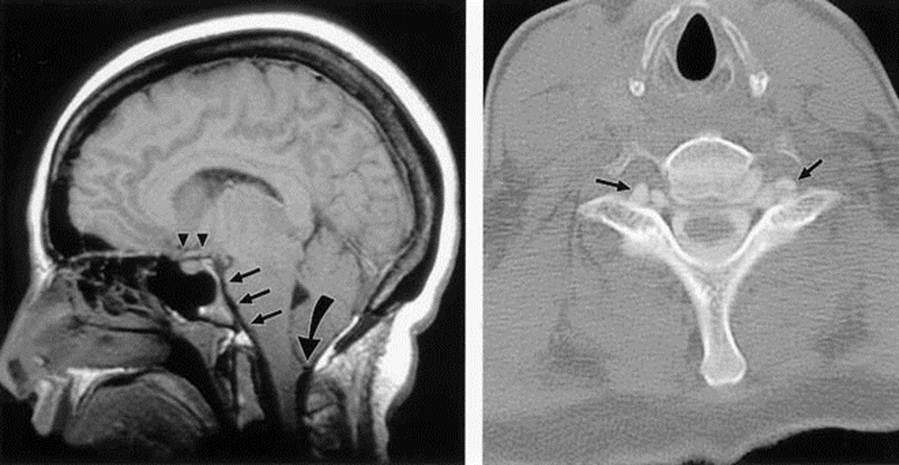
– Reproduced with permission from Marcel Maya, MD and Cedars-Sinai Medical Center, Los Angeles, CA –
If Chiari is seen on imaging, usually MRI, then reversible causes such as intracranial hypotension from spinal CSF leaking must be ruled out. In cases of intracranial hypotension, additional imaging findings will usually be present, which will help with correct diagnosis.
The imaging findings on cranial MRI in cases of intracranial hypotension include:
S = subdural fluid collections
E = enhancement of pachymeninges
E = engorgement of venous structures
P = pituitary hyperemia
S = sagging of brain
Remember, Chiari can be congenital or acquired. An underlying cause such as intracranial hypotension should be considered so that the most appropriate treatment can be planned.