

The initial treatment of spontaneous spinal cerebrospinal fluid (CSF) leaks does not require localization of the CSF leak with spinal imaging, since one or more non-directed epidural blood patches are often effective. In those situations where conservative measures or non-directed epidural blood patches fail to result in resolution of symptoms, spinal imaging is used to localize the CSF leaks for targeted treatment. Accuracy of findings can impact outcomes.
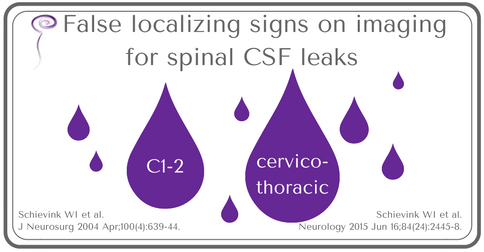
These authors have previously reported that contrast seen at cervical levels 1 and 2 is a false localizing sign. This paper discusses another area of discordance between imaging findings and operative findings, demonstrating that myelographic evidence of contrast at the cervico-thoracic junction is often another false localizing sign.
This study reports on 13 patients (9 women and 4 men) who presented with orthostatic headache and myelographic imaging findings of extradural contrast along nerve roots at the cervico-thoracic junction. All patients had epidural patching procedures but later went on to have surgery. CSF leak at the cervico-thoracic junction was confirmed at this level in only 2 of the patients at surgery. Nine of the remaining 11 patients later went on to have further imaging, specifically digital subtraction myelography. In all 9 patients, the site of the CSF leak was found to be at a thoracic level, not at the cervico-thoracic junction.
Physicians treating patients should be aware of both false localizing signs on imaging: C1-2 and cervicothoracic levels because these can lead to misdirected treatment.
As an increasing number of patients with spontaneous intracranial hypotension secondary to CSF leaks are evaluated and treated, imaging techniques will continue to be refined and impact treatment plans and outcomes favorably.
Here is the recent abstract:
False localizing sign of cervico-thoracic CSF leak in spontaneous intracranial hypotension
Schievink WI, Maya MM, Chu RM, Moser FG.
Neurology. 2015 Jun 16;84(24):2445-8.
Abstract
OBJECTIVE:
Spontaneous spinal CSF leaks are an important cause of new-onset headaches. Such leaks are reported to be particularly common at the cervico-thoracic junction. The authors undertook a study to determine the significance of these cervico-thoracic CSF leaks.
METHODS:
The patient population consisted of a consecutive group of 13 patients who underwent surgery for CSF leak repair based on CT myelography showing CSF extravasation at the cervico-thoracic junction but without any evidence of an underlying structural lesion.
RESULTS:
The mean age of the 9 women and 4 men was 41.2 years. Extensive extrathecal longitudinal CSF collections were demonstrated in 11 patients. At surgery, small leaking arachnoid cysts were found in 2 patients. In the remaining 11 patients, no clear source of CSF leakage could be identified at surgery. Resolution of symptoms was achieved in both patients with leaking arachnoid cysts, but in only 3 of the 11 patients with negative intraoperative findings. Postoperative spinal imaging was performed in 9 of the 11 patients with negative intraoperative findings and showed persistence of the longitudinal intraspinal extradural CSF. Further imaging revealed the site of the CSF leak to be ventral to the thoracic spinal cord. Five of these patients underwent microsurgical repair of the ventral CSF leak with resolution of symptoms in all 5 patients.
CONCLUSIONS:
Cervico-thoracic extravasation of dye on myelography does not necessarily indicate the site of the CSF leak. Treatment directed at this site should not be expected to have a high probability of sustained improvement of symptoms.
PMID: 25979700
neurology.org/content/early/2015/05/15/WNL.0000000000001697.abstract
Here is the earlier paper abstract:
False localizing sign of C1-2 cerebrospinal fluid leak in spontaneous intracranial hypotension.
Schievink WI1, Maya MM, Tourje J.
J Neurosurg. 2004 Apr;100(4):639-44.
Abstract
OBJECT:
Spontaneous intracranial hypotension due to a spinal cerebrospinal fluid (CSF) leak is an important cause of new daily persistent headaches. Spinal neuroimaging is important in the treatment of these patients, particularly when direct repair of the CSF leak is contemplated. Retrospinal C1-2 fluid collections may be noted on spinal imaging and these are generally believed to correspond to the site of the CSF leak. The authors undertook a study to determine the significance of these C1-2 fluid collections.
METHODS:
The patient population consisted of a consecutive group of 25 patients (18 female and seven male) who were evaluated for surgical repair of a spontaneous spinal CSF leak. The mean age of the 18 patients was 38 years (range 13-72 years). All patients underwent computerized tomography myelography. Three patients (12%) had extensive retrospinal C1-2 fluid collections; the mean age of this woman and these two men was 41 years (range 39-43 years). The actual site of the CSF leak was located at the lower cervical spine in these patients and did not correspond to the site of the retrospinal C1-2 fluid collection.
CONCLUSIONS:
A retrospinal fluid collection at the C1-2 level does not necessarily indicate the site of the CSF leak in patients with spontaneous intracranial hypotension. This is an important consideration in the treatment of these patients because therapy may be inadvertently directed at this site.
PMID: 15070118